Coracobrachialis – An Overview
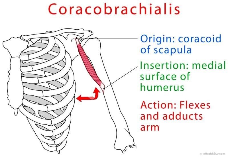
The coracobrachialis, also known as the rotator cuff, is a small, thin muscle that originates behind the coracobrachialis (the white area on the shoulder). The tendon muscle runs inferolaterally to the sternum. They insert onto the coracoderm’s anteroposterior surface, near the acromion. The tendon is found generally between the front of the clbumere, and the anterior part of the acromion.
Functions
The primary function of the coracobrachialis, is to provide stability and strength to the inferior and middle face of the clavicle, while preventing it from flexing and collapsing during prolonged standing and other activities. The insertion into the coracobrachialis helps increase blood supply to the arm by extending the branches of the brachial artery, thereby allowing for more stable blood flow. As the amount of blood supply increases, the amount of internal force applied to the arm decreases. This leads to less tension in the tendons and muscles of the upper arms.
Causes
The acromion process, also known as coracobrachiosaphragmatic separation, is caused by the acromion, which is caused by the insertion of a nerve from the upper cervical spine to the forearm, or from the paraspinal nerves to the forearm. The nerve, called the inguinal nerve, inserts onto the coracobrachialis gliding along the superior border of the coracobrachialis and extends to the abdominal wall and then onto the inferior frontal wall. This process is called “nutcracking.” Other causes of this condition are: injuries, inflammation, infection, and tumors.
If there is a weakness in the muscles around the coracoacromial ligament, the coracobrachialis may separate and become inflamed. Common causes of the inflammation include injury, infection, and tumors. The inflammation can be very severe, resulting in the formation of scar tissue and loss of joint mobility.
The humerus is the major muscle in the body responsible for transmitting power from the forearm to the rest of the body. This muscle connects the medial surface of the coracobrachialis to the rest of the arm. There are two major types of humerus: the deep and shallow. The deep humerus is extremely rare, while the shallow humerus is extremely common.
Median Nerve
The nerves that enter the coracobrachialis are the nerves that provide the arms with movement. The most common nerves entering the coracobrachialis are the median nerve, the ulnar nerve, and the nerve. Injury to any of these nerves can result in weakness or even complete loss of arm movement. Infection, inflammation, and tumor growth can also result in severe problems. The ulnar nerve and Testicularis nerve are the major nerves that run through the entire arm.
Abnormal Content & Nerve Connections
The abnormal content published on the coracobrachialis muscle can affect the nerve connections of this structure. In the most common situation, the abnormal content published on the coracobrachialis muscle causes a complete loss of arm movement. Treatment of this condition generally requires an arthroscopic procedure that removes the diseased material from the muscle. While this method is effective, it does not cure the problem.
Abnormal Content & Median Nerve
The abnormal material that is published on the coracobrachialis muscle can also affect the nerve connections of the median nerve that runs through the middle section of the arm. This disease can cause weakness and numbness in the thumb, index finger, middle and index fingers as well as the palm side of both the thumbs. Treatment of this condition generally requires surgery that removes the diseased tissue from the muscle. A Tendonectomy, however, is another option for treatment. Tendonectomies are performed by sliding the ends of a tendon inward, which reduces the amount of motion of this muscle.
The second structure affected by this disorder include the jugular bulb and the internal capsule. This disorder affects the function of the jugular bulb, which supplies blood to the brain and eyes. The internal capsule is the key structure that separates the spleen from the rest of the body. An injury to this structure can cause weakness, nausea, vomiting, and severe pain. The pathology has many similarities to that of cervical spondylosis, which is an injury to the neck and head section of the spine.
Medius & Brevis
Medius and brevis refer to the muscles that cover the lower half of the abdomen. The abbreviation medius refers to the muscle that wraps around the upper abdominal wall. The abbreviation brevis refers to the lower abdominal wall that wraps around the lower torso. The three parts of the coracoid process are the medulla, the peritoneum, and the tunica. The structure of the three sections is important in diagnosing this disease since they each contribute to the pathophysiology.
Diagnosis
The diagnosis of Cervical Spondylosis involves the palpitation of the neck and the two sides of the head. If it is detected in the early stages, it can be treated with conservative treatments. When the disease has reached a certain level, invasive procedures such as a lumbar laminectomy and coracoacromial ligament repair are required. If the Cervical Spondylosis has spread beyond the peritoneum and lamina, it will require open surgery for complete removal of the affected vertebrae.