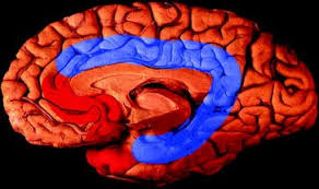
Cingulate Gyrus and Its Linked to Emotions?
The cingulate gyrus is situated above the parietal cortex and forms a part of the temporal limbic network. Cingular seizures are characterized by seizures, which are characterized by the sudden, temporary loss of muscular coordination and movement co-ordination. It is often accompanied by visual artifacts and by feelings of unreality or derealization. Cingular seizures can cause a wide array of different clinical presentations, which correlate with the underlying anatomic and structural subdivisions of the cingulate cortisone complex.
The parts of the brain which are affected by seizures are the left parietal cortex (medial temporal), the right parietal cortex (middle temporal), and the lateral portion of the cerebellum. A disconnection between these three regions results in a disruption of memory and the related functions of the brain. The parts of the brain which are affected by seizures are not completely understood, although research has shown a considerable correspondence between the cognitive and emotional aspects of anxiety and depression. The parts of the brain which are affected by seizures include the amygdala, hippocampus, periaqueductal grey matter (PAG), and basal ganglia. There is also considerable correspondence between the emotional aspects and behaviour of anxiety and depression and abnormalities of the cingulate gyrus.
The major functions that are affected by seizures are: speech, language, memory, judgment, attention, movement, facial expression, vision, smell, taste, and chewing. The major functions, which are not affected by the disorder include movement, thought, sensation, and movement. This is because the human brain cannot easily distinguish between normal activity, abnormal activity, or signals that are not pertinent to an individual state. As such, the cingulate gyrus participates in the integration of external stimuli within the context of the individual’s context.
The two major portions of the brain, the frontal cortex and the posterior cingulate gyrus, are directly attached to each other and the location of their exact locations is highly related to the function that they perform. There is a junction of the frontal cortex with the prefrontal cortex and the superior parietal region. In addition to this, there is a junction of the corticospinal tract and the thalamus. These areas of the brain are intimately connected with each other and they form a network which is responsible for the generation of sensory inputs and the transmission of motor information across the entire cortisone mediated limbic system.
The connections between the front lobes and the posterior cingulate gyrus arise through the reciprocal connections which exist between the two regions of the brain. These reciprocal connections are established through the projection of nerve fibers from the frontal cortex to the thalamus. The thalamus then projects its impulses to the various areas of the body which it controls. This is an extremely complex process that is only understood fully by the science of neurology. However, the basic functioning of this process remains unaltered when the corpus callosum is present. The corpus callosum optimally connects all the parts of the brain that have a common pathway through the nucleus accumbens.
Neuromuscular diagrams and neuropsychologists have been used for many years to determine the location and the function of the different parts of the human brain. Through the years, the method of performing a cingulate gyrus analysis has evolved into a much more efficient method in terms of cerebral anatomy and function diagnosis. In most cases, this type of analysis is accomplished through the use of magnetic resonance imaging (MRI) and the placement of one of three types of electrodes: the magnetized shunt coil technique, the periemagic technique or the transverse electronics technique. Each of these methods is effective at different times, however, it all boils down to a particular question of the function: where is the medial surface of the brain?
In cases where the information from MRI and/or PET scans show strong evidence for the presence of abnormalities in the structure of the brain, it may be determined that a section of the cingulate gyrus has not completely developed. The term “cringate herniation” is commonly used by neurologists to describe a situation where portions of the cerebral hemispheres are missing. The condition is often associated with progressive cerebellar ataxia, and symptoms such as seizures, speech regression, ataxia, hypotonia, ambience, and visual illusions may occur. Other conditions that have similar clinical manifestations are paired claudication, progressive cerebellar ataxia, stroke, hemiplegia or hemiparesis, bradykinesia, oculomotor problems and many others.
Subregions of the cingulate gyrus are indeed important for the well-being of humans. Nevertheless, the existence of a large section of the brain that does not develop or fully mature during the developmental process has some disturbing implications for the way humans think, feel, and behave. A dysfunctional subgroup of the cingulate gyrus has been linked with psychiatric disorders such as depression, schizophrenia and other psychological conditions. More importantly, there is convincing evidence that the condition is closely related to emotional states. In a number of recent functional imaging studies, caregivers of patients with an autism spectrum disorder have revealed that emotional states can be reflected in brain regions that are typically not activated during behavioral tasks.